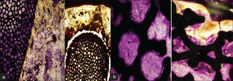
Figure 4.

(a) 12.5× image showing well formed bony trabeculae in contact with implant surface without gap (Lt). Cancellous bone around tip is not new bone formation (Rt). (b) 100× image showing implant pores with active proliferation of young fibroblasts in vascular rich stroma and dense celullar rim lining the surface of implant material (Rt). (The cellular rim is supposed to be a possible osteoblastic proliferation that could not be technically evaluated in specimen.) Several foci of ingrowing new bone into porous implant sprouting from interface zone were evident (left, arrow)
