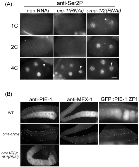
Fig. 2.
OMA-1/2 repress transcription in P2 indirectly by preventing PIE-1 degradation. (A) Anti-Ser2P staining in 1-cell, 2-cell and 4-cell embryos of wild-type or indicated RNAi animals. Arrowhead indicates the P2 nucleus. (B) Immunofluorescence micrographs of anti-PIE-1, anti-MEX-1 and GFP fluorescence micrograph of GFP::PIE-1 ZF1 in the gonads (dashed outline) of wild-type, oma-1(te33);oma-2(51) or oma-1(te33);oma-2(te51);zif-1(RNAi) animals. Scale bars: 10 μm in A; 30 μm in B.

