Fig. 4.
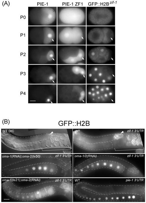
GFP::H2Bzif-1 recapitulates the temporal and spatial localization of ZIF-1 activity. (A) Fluorescence micrographs of staged embryos expressing indicated GFP reporters. Embryo stage is indicated to the left by the name of its germline blastomere (arrow). (B) GFP::H2B expression from reporters containing indicated 3′ UTR (upper right hand corner) in gonads of different genetic backgrounds (upper left hand corner). A wild-type gonad exhibiting GFP::H2Bzif-1 in mitotic germline stem cells (arrowheads), but repressed in meiotic germ cells, is shown with both DIC (top left) and fluorescence micrographs (top right). GFP::H2B expressed from another reporter construct differing only in the 3′ UTR is expressed in oocytes (bottom right). Additional data demonstrating specificity of GFP::H2Bzif-1 is shown in Fig. S3 in the supplementary material. Scale bars: 10 μm in A; 30 μm in B.

