Figure 3.
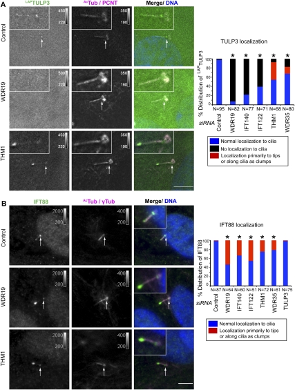
TULP3 localizes to the primary cilium dependent on the IFT-A complex. (A) RPE LAPTULP3 line A cells were transfected with siRNAs (indicated to the left of the micrograph panels) for 72 h and serum-starved for the last 24 h before fixing and staining for pericentrosomal pericentrin (PCNT, magenta), the axonemal marker Ac-tubulin (AcTub, magenta), and DNA (blue). White arrows indicate the ciliary base. Bars, 5 μm. Quantification of localization of LAPTULP3 to the cilia in similar assays is shown to the right. (*) P < 0.0001 with respect to control, using a χ2 test. (B) RPE cells were transfected with the indicated siRNAs for 72 h and serum-starved for the last 24 h before fixing and staining for IFT88 (green), pericentrosomal γ-tubulin (γTub, magenta), the axonemal marker Ac-tubulin (AcTub, magenta), and DNA (blue). White arrows indicate the ciliary base. Bars, 5 μm. Quantification and statistical significance are as in A.

