Figure 5.
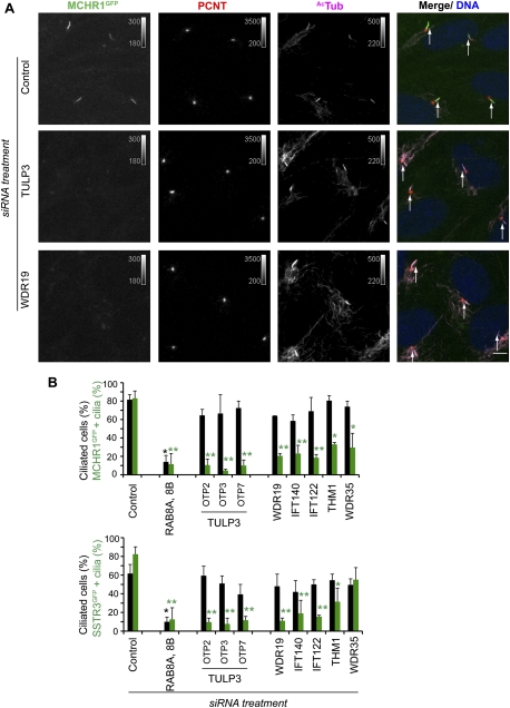
TULP3 and IFT-A coregulate localization of ciliary GPCRs. (A) RPE MCHR1GFP stable cells were transfected with the indicated siRNAs for 78 h and serum-starved for the last 30 h before fixing and staining for pericentrin (PCNT, red), Ac-tubulin (AcTub, magenta), and DNA (blue). White arrows indicate cilia. Bars, 5 μm. (B) Percentages of total cilia and GFP-positive cilia in assays similar to A were counted in two to three independent experiments with RPE MCHR1GFP and RPE SSTR3GFP stable cells. Error bars represent SEM. (*) P < 0.05; (**) P < 0.001 with respect to control in each group. See also Supplemental Figure S5.

