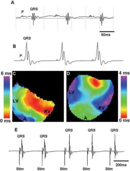
Figure 4.
Class II mutants exhibited intermittent use of bypass tracts. A, Surface ECG of a class II mutant showed normal AV conduction. B, vECG of the same heart after isolation and Langendorff perfusion clearly demonstrated conduction through a bypass tract. C and D, Activation maps showed bypass conduction (C) followed by a collision beat (D) where signals were coming both from the apex and the base of the ventricle in a single heart. E, vECG data from the same heart demonstrated intermittent use of the bypass during pacing. First 2 beats had a short PR interval, which indicates accessory pathway conduction, whereas the next 3 beats were conducted through the AV node. P indicates atrial depolarization; QRS, ventricular depolarization; LV, left ventricle; RV, right ventricle; A, apex; and Stim, stimulus.

