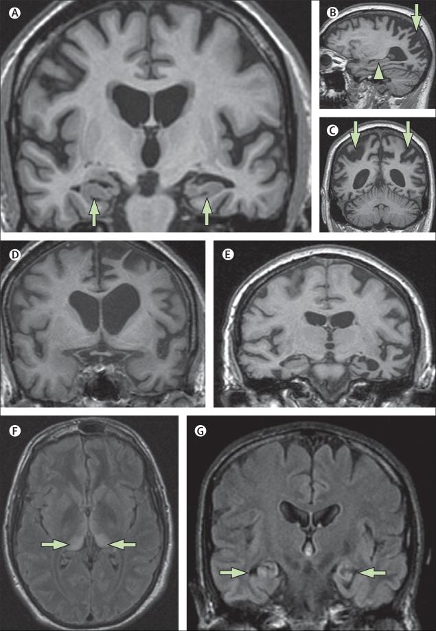
Figure 4. The value of MRI in investigation of young-onset dementia.
(A) Mild Alzheimer's disease in a 60-year-old individual with sporadic Alzheimer's disease (T1-weighted MRI): atrophy of hippocampi (arrows) is the earliest feature in amnestic Alzheimer's disease but hippocampi might appear normal, particularly in younger patients with Alzheimer's disease. (B, C) Posterior cortical atrophy in a 58-year-old individual (T1-weighted MRI). The sagittal view (B) shows a relatively well preserved hippocampus (arrow head); parieto-occipital atrophy (arrows) is seen on sagittal (B) and coronal (C) views. (D) T1-weighted MRI of a 58-year-old individual with progressive non-fluent aphasia who had pathologically proven Pick's disease. (E) T1-weighted MRI of a 63-year-old woman with semantic dementia who had tau-negative, TARDBP-positive inclusions at autopsy. (F) A 21-year-old individual with increased signal bilaterally in the pulvinar (arrows) on axial FLAIR MRI. The pulvinar sign is indicative of variant Creutzfeldt-Jakob disease and is best seen on axial FLAIR (or T2-weighted) MRI where the postero-medial thalami are brighter than the basal ganglia. Variant Creutzfeldt-Jakob disease was subsequently confirmed on tonsillar biopsy. (G) By use of FLAIR MRI, bilateral hippocampal high signal and atrophy is shown in a 57-year-old man with voltage-gated potassium channel antibody limbic encephalitis (arrows). FLAIR=fluid attenuated inversion recovery. TARDBP=TAR-DNA binding protein (also known as TDP-43).

