Fig. 3.
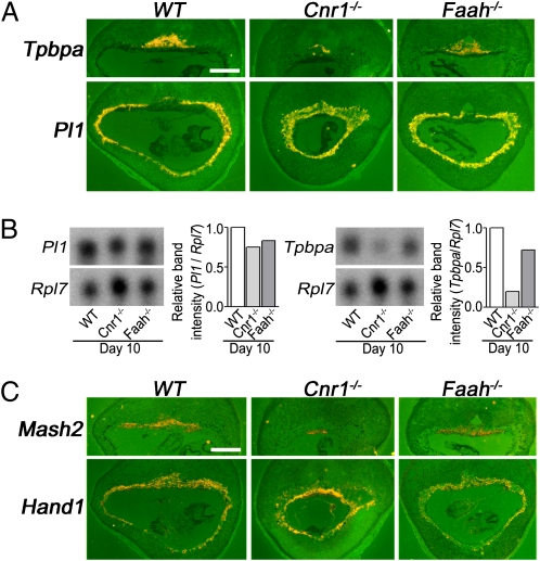
Cnr1−/− spongy layer is developmentally compromised. (A) Although the number of Tpbpa positive cells is considerably lower in Cnr1−/− placentas compared with WT and Faah−/− placentas, Pl1 expression is similar in placentas of all three genotypes on day 10. (Scale bar, 1,000 μm.) (B) Northern blot analyses of WT, Cnr1−/−, and Faah−/− implantation sites on day 10 of pregnancy show that Tpbpa, but not Pl1, levels in Cnr1−/− mice are reduced. Levels of Tpbpa and Pl1 mRNAs were normalized and quantified against Rpl7 (a house-keeping gene) using the same membrane. (C) Levels of mash2 transcripts were reduced in Cnr1−/− placentas, but Hand1 levels were comparable in WT, Cnr1−/−, and Faah−/− placentas. (Scale bar, 1,000 μm.)

