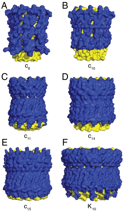
Fig. 3.
External surfaces of rotor rings from F- and V-ATPases. The rings are shown in solid representation. The N- and C-terminal α-helices are yellow and blue, respectively. Parts A–E, c-rings from F-ATPases from bovine and yeast mitochondria, from Ilyobacter tartaricus, from Spinacea oleracea, and from Spirulina platensis. Part F, the K-ring from the V-ATPase from Enterococcus hirae.

