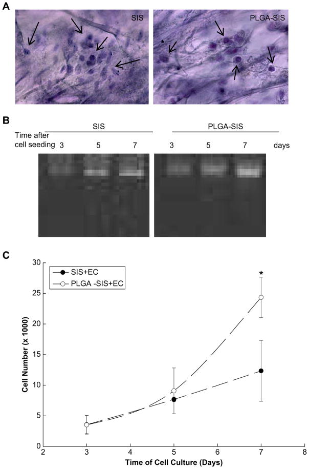
Fig. 5.
Endothelial cell growth on PLGA NP modified SIS. (A) Morphological presentation of HEMC-1 cells on unmodified SIS and PLGA NP modified SIS. Arrows indicate the presence of cells in SIS structure. (B) HEMC-1 proliferation on SIS as determined by genomic DNA contents was presented over a period of 7 days following cell seeding. (C) Quantitative presentation of HEMC-1 growth on SIS. Results were presented as mean ± standard deviation of cell numbers from three independent experiments. * Indicates the results were different vs. unmodified SIS on day 7 (p < 0.05).

