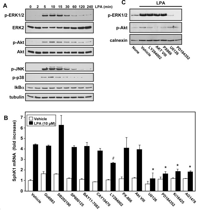
Fig. 2. Effects of inhibitors of LPA signaling pathways on SphK1 upregulation.
(A) MKN1 cells were stimulated with 1 μM LPA for the indicated times, equal amounts of cell lysates were separated by SDS-PAGE and analyzed by immunoblotting with the indicated antibodies. Blots were stripped and immunoblotted with anti-ERK2, anti-Akt, and anti-tubulin to ensure equal loading and transfer. (B) MKN1 cells were pretreated with vehicle or with Ki16425 (10 μM), AG1478 (1 μM), Go6983 (10 μM), SB202190 (1 μM), SP600125 (10 μM), BAY11-7082 (1 μM), CAY10470 (1 μM), LY294002 (10 μM), PX-866 (100 nM), Akt VIII (10 μM), U0126 (10 μM), or PD184352 (10 μM), for 1 hour, and subsequently stimulated with LPA (10 μM) for 6 hours. RNA was isolated, reverse-transcribed, and SphK1 mRNA measured by QPCR. Data are expressed as fold induction after normalization to GAPDH. *, P < 0.001, #, P< 0.05 compared to LPA treated cells. (C) Cultures were pretreated with the indicated inhibitors for 1 hour prior to stimulation with LPA (10 μM) for 20 min. Equal amounts of lysate proteins were analyzed by immunoblotting with anti-phospho-Akt and anti-phospho-ERK1/2 antibodies. Blots were stripped and reprobed with anti-calnexin antibody to ensure equal loading and transfer.

