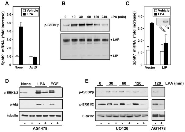
Fig. 4. Involvement of C/EPBβ in LPA-induced transcriptional upregulation of SphK1.
(A) MKN1 cells were pretreated for 1 hour with actinomycin D (5 μg/ml) and then stimulated for 6 hours without or with LPA and SphK1 mRNA measured by QPCR. (B) Cells were stimulated with LPA for the indicated times and equal amounts of lysates analyzed by immunoblotting with anti-phospho-C/EPBβ (Thr235) antibody or with antiC/EPBβ antibody. The LAP and LIP subunits are indicated. (C) MKN1 cells transfected with vector or LIP were stimulated without or with LPA for 6 hours and SphK1 mRNA measured by QPCR (Insert: western blotting demonstrating LIP expression). (D,E) MKN1 cells were pretreated for 30 min with vehicle, AG1478 (1 μM) (D,E), or U0126 (10 μM) (E), as indicated. Cells were then stimulated for 5 min with vehicle, LPA (10 μM), or EGF (10 ng/ml) (D), or for the designated times (E), as indicated. Proteins were analyzed by immunoblotting with antibodies against phospho-ERK1/2, phospho-Akt, tubulin, phospho-C/EPBβ (Thr235), or ERK1/2.

