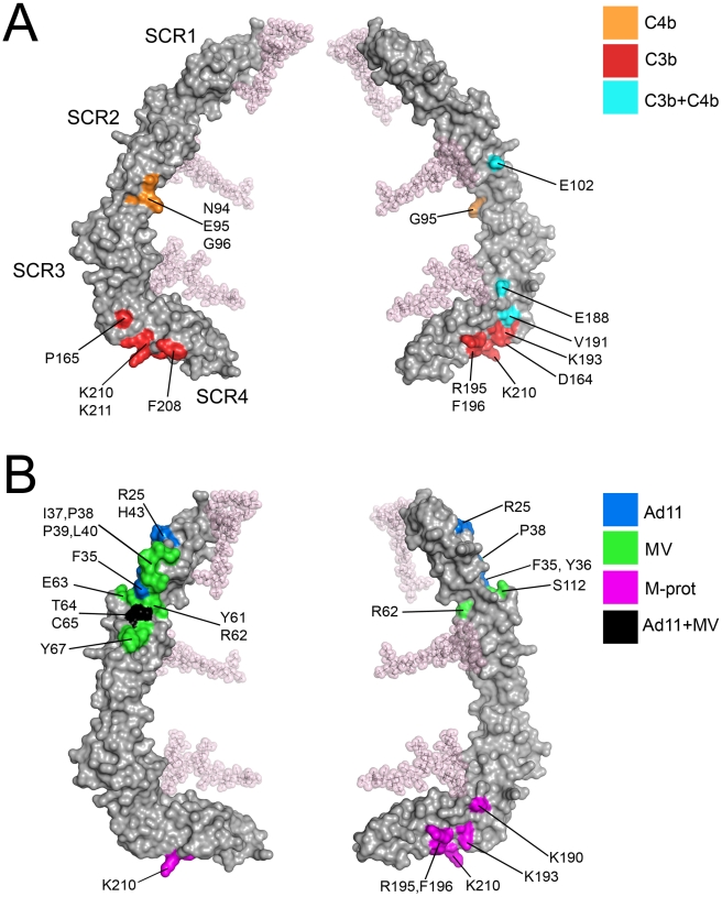
Figure 6. Ligand binding surfaces in the CD46-4D protein.
Two views of the CD46-4D structure (grey), differing by 180 degrees along a vertical axis, are shown in each case. (A) Surface representations of CD46-4D, with regions implicated in C3b- (red), C4b- (orange) and C3b + C4b-binding (blue) shown in color [35], [36], [37]. Individual residues are indicated. (B) Surface representations of CD46-4D, with regions known to interact with Ad11 and MV [34] shown in blue and green, respectively. Regions that interact with both viruses are highlighted in black. Residues predicted to contact the Streptococcus M protein (M-prot) [38] are shown in purple.

