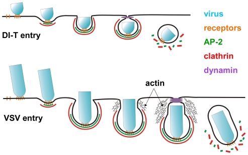
Figure 7. Model of DI-T and VSV entry.
DI-T (above) and VSV (below) particles engage host cells through interactions between the viral surface glycoproteins and unknown cellular receptor moieties. Following attachment, both particle types undergo slow diffusion (diffusion coefficient ∼5×10⁻¹¹ cm2 s−1) on the cell surface for an average of ∼2 min. before being captured by a clathrin-coated pit. For DI-T, continued clathrin assembly drives complete particle envelopment by the plasma membrane and leads to virus endocytosis. In contrast, the presence of a VSV particle in a coated pit physically prevents complete membrane constriction by the clathrin machinery and causes clathrin assembly to halt prematurely. The force provided by actin polymerization then further remodels the plasma membrane and thereby encloses the virus particle in a partially clathrin-coated vesicle.

