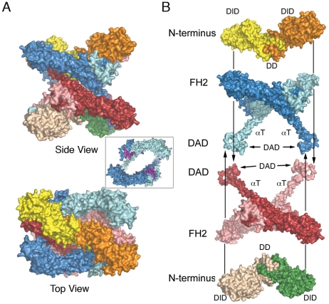
Figure 4. Surface views of the autoinhibited mDia1 N+C tetramer.
A, Side and top views of the tetramer, colored as in Figure 1B. Inset: top view of one FH2 dimer, with the residues expected to bind actin shaded magenta. Note that packing of the N-terminal DID-DD dimers onto the FH2 domain blocks the actin-binding surface. B, “Exploded” view depicting construction of the tetramer.

