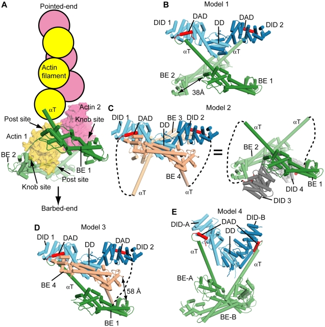
Figure 3. Structural models for mDia1 autoinhibition.
(A) A structural model of the FH2 domain at a filament barbed-end is presented, in which a cartoon actin filament is placed at the pointed-end side of the crystal structure of Bni1p FH2 domain bound to TMR-actin (PDB code: 1Y64). (B-E) Structural models of the mDia1 (DID-DD)2•(FH2-DAD)2 dimer derived from the crystal. Dashed lines indicate linkers connecting the bridge elements and DADs. For clarity, the invisible linkers connecting two bridge elements in each model are not indicated. The names and numbering for each structural element in this figure are same as those in Figure 2 except (E) Model 4, in which two DIDs are labeled as DID-A and DID-B and the bridge elements as BE-A and BE-B. (B) In Model 1, no swapping of αT is assumed. This leads to only one bridge element bound to each DID. The dotted line is a 5 residue linker missing in the electron density map. In this configuration, the distance spanned between the two bridge elements by the lasso (∼30 residues not observed in the electron density) is 38 Å, as indicated in the figure. (C) For Model 2, two identical models are shown for clearer presentation. The model on left possess the same (DID-DD)2 dimer as in (B) and (D). The right is composed of the other pair from the tetramer and is to facilitate comparison with (A). The distance between the ends of dotted lines (Met1172 and Thr1179) is 73 Å, which is too long for the missing linker (5 residues) to connect. This operation would require at least the C-terminal 3∼5 turns of αT to melt. (D) Model 3 is a hybrid model between Models 1 and 2, in which αT of BE4 is swapped. The distance spanned by the missing portion of the lasso is 58 Å, as indicated in the figure. (E) In Model 4, the DID•BE pairs in Model 1 were organized according to the asymmetric structure of free DID-DD (2BNX). Thus, the relative positions within each DD-DID•DAD-BE element are the same as in (B), i.e., the position of BE-A relative to DID-A, BE-B to DID-B (Model 4), BE1 to DID1, and BE2 to DID2 (Model 1) are all the same. However, the different organization of the two DIDs in Model 1 and the asymmetric (DID-DD)2 dimer results in a different organization of the BEs in the two cases.

