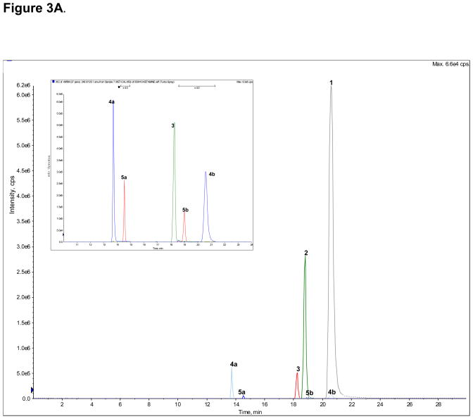
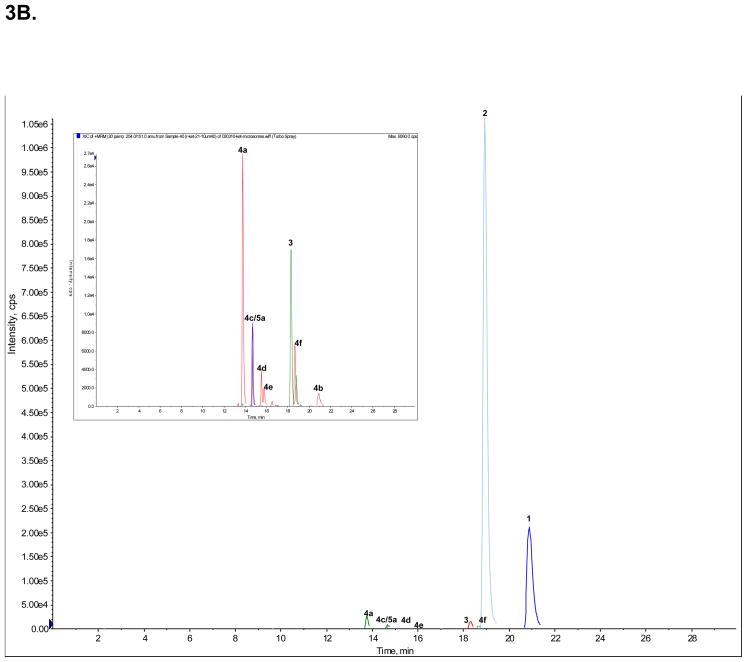
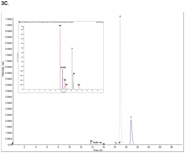
Figure 3.
The achiral separation of (R,S)-Ket (peak 1), (R,S)-norKet (2), (R,S)-DHNK (3), (2S,6S;2R,6R)-HNK (4a), (2S,6R;2R,6S)-HNK (4b), (2S,6S)-HKet (5a) and (2S,6R)-HKet (5b) on a C18 column, where: A. Standard solution; B. Extracted sample obtained from the incubation of (R)-Ket with human microsomal preparation; C. Extracted sample obtained from the incubation of (S)-Ket with human microsomal preparations. See Table 2 for the assignment of the structures for peaks 4c–f, 5c–d and 6a–e and the text for experimental details.


