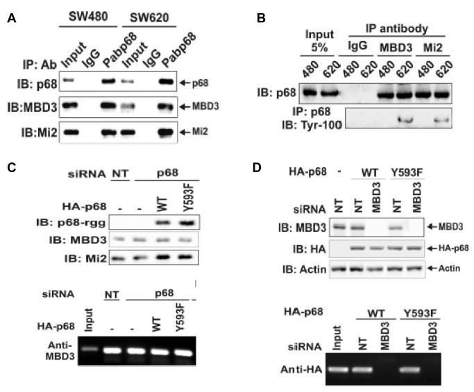
Figure 3.
P68 interacted with the MBD3:Mi-2/NuRD complex.
(A) Co-IPs of MBD3 and Mi-2 with p68 in SW480 and SW620 cells were detected by IB of p68 co-immunoprecipitates using appropriate antibodies (indicated). P68 was precipitated by polyclonal antibody Pab-p68. Rabbit IgG was used as a negative control IP antibody. Inputs were the IBs of extracts without IP. (B) Co-IPs of p68 with MBD3 and Mi-2 in SW620 (620) and SW480 (480) cells were detected by IBs of co-immunoprecipitates of antibodies (anti-MBD3 and anti-Mi-2) using monoclonal antibody p68-rgg. Mouse IgG was used as control IP antibody. The inputs were the IBs of extracts without IP. The tyrosine phosphorylation of p68 was detected by IB of PAbp68 immunoprecipitated p68 using antibody Tyr-100. (C) Upper panel, cellular levels of MBD3, HDAC1, and exogenously expressed HA-p68s (WT or Y593F mutant) were analyzed via IBs using appropriate antibodies (indicated). The IBs were performed with cellular extracts made from SW620 cells that were treated with p68 siRNA (p68) or non-targeting siRNA (NT). Lower panel, Interactions of MBD3 with Snail1 promoter in the cells treated as described in upper panels were detected by ChIP assays using anti-MBD3 antibody (Anti-MBD3). (D) Upper panel, cellular levels of MBD3 and exogenously expressed HA-p68s (WT or Y593F mutant) (without endogenous p68 knockdown) were analyzed via IBs using appropriate antibodies (indicated). The IBs were performed with cellular extracts made from SW620 cells that were treated with MBD3 siRNA (MBD3) or non-targeting siRNA (NT). IB of Actin was a loading control. Lower panel, Interactions of HA-p68 with Snail1 promoter in the cells treated as described in upper panels were detected by ChIP assays using anti-HA antibody (Anti-HA).

