Figure 5.
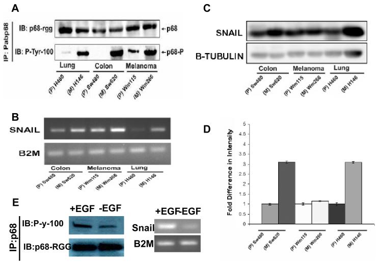
P68 phosphorylation correlates with Snail1 expression in metastatic and non-metastatic cancer cells.
(A) P68 tyrosine phosphorylation in three pair of cancer cell lines (indicated). The tyrosine phosphorylation of p68 was detected by immunoblot (via the anti-phosphor-tyrosine antibody; IB:P-tyr-100) of p68 that was immunoprecipitated from nuclear extracts of the cells using the antibody Pabp68 (IP:Pabp68). Immunoblot of p68 in the IPs using the antibody p68-rgg (IB:p68- rgg) was the loading control. (B) The expression levels of Snail1 mRNA in the three pair of cancer cells (indicated) were detected by RT-PCR of total RNAs isolated from the cells. The RT-PCR detections of mRNA of B2M (B2M) gene in the cells were the controls. (C) The cellular Snail1 protein levels in the three pair of cancer cells (indicated) were examined by immunoblot of the cell lysate prepared from the cells using antibody against Snail1 (IB:Snail1). IB of β-tubulin was used as the loading control. (D) is the quantification (average) of the immunoblot signals of Snail1 after normalizing to the loading control β-tubulin blots. The error bars represent standard deviation of results from three independent immunoblots. (E) (Left) Tyrosine phosphorylation of p68 in SW480 cells that were treated (+EGF) or untreated (−EGF) with EGF (25 ng/ml) was analyzed by Immunoprecipitation of p68 (IP:p68) from the cell lysates followed by immunoblot using the antibody P-y-1oo (IB:P-y-100). Immunoblot of p68 (IB:p68-RGG) indicated the precipitated p68. (Right) Expression of Snail1(Snail) in SW480 cells that were treated (+EGF) or untreated (−EGF) with EGF (25 ng/ml) was analyzed by RT-PCR of the total RNA isolated from cell lysates. RT-PCR analyses of B2M mRNA in the EGF treated or untreated cells is a control.

