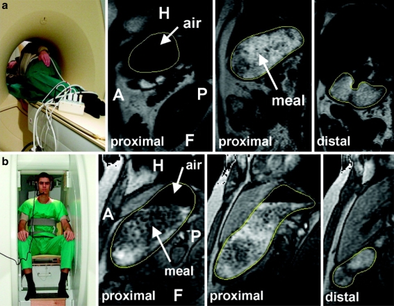
Fig. 4.
a T2-weighted coronal slice-selective images of subject 1 depicting the stomach in its full J-shaped form. For dynamic study, respiratory gating determined data acquisition at times 0, 3.2, 6.5, 9.5 and 13.3 s. The contractions started in the proximal part of the body and moved aborally toward the antrum. The mean velocity was approximately 49.5 cm/min. b Transaxial scan through the middle part of the stomach. Nearly symmetrically and concentrically configured constrictor rings can be assessed. The in-plane resolution of 1.4 mm × 1.4 mm was sufficient to identify the longitudinal fissure of the gastric wall

