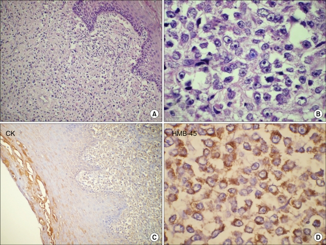
Fig. 1.
Paraffin section of cervical growth (A), showing diffuse sheets of round to spindle shaped undifferentiated malignant cells with focal epitheliotropism (H&E, ×100). High power (B) showing few binucleate and multinucleate bizarre tumor cells with prominent eosinophilic nucleoli (H&E, ×400). No pigment was seen within the tumor. Tumor cells were negative for cytokeratin (C, streptavidin-biotin immunoperoxidase CK, ×100) and show strong immunoreactivity for HMB-45 antigen (D, streptavidin-biotin immunoperoxidase HMB-45, ×400).

