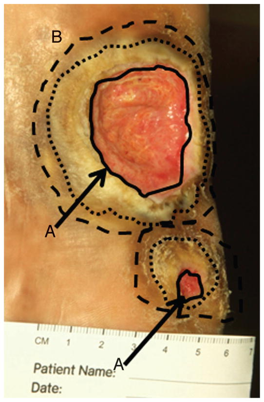
Figure 4. BIOLOGICALLY BASED MARGINS OF DEBRIDEMENT.
Digital photograph of a neuropathic plantar foot ulcer in a patient with diabetes. The solid black lines indicate nonhealing edge (location A, indicated by black arrows). Two outer circles (broken lines) indicate 2 possible margins of debridement, the presumed location B or healing-edge of the wound. At this edge, keratinocytes have the ability to migrate and participate in the wound healing process.

