
Figure 1.
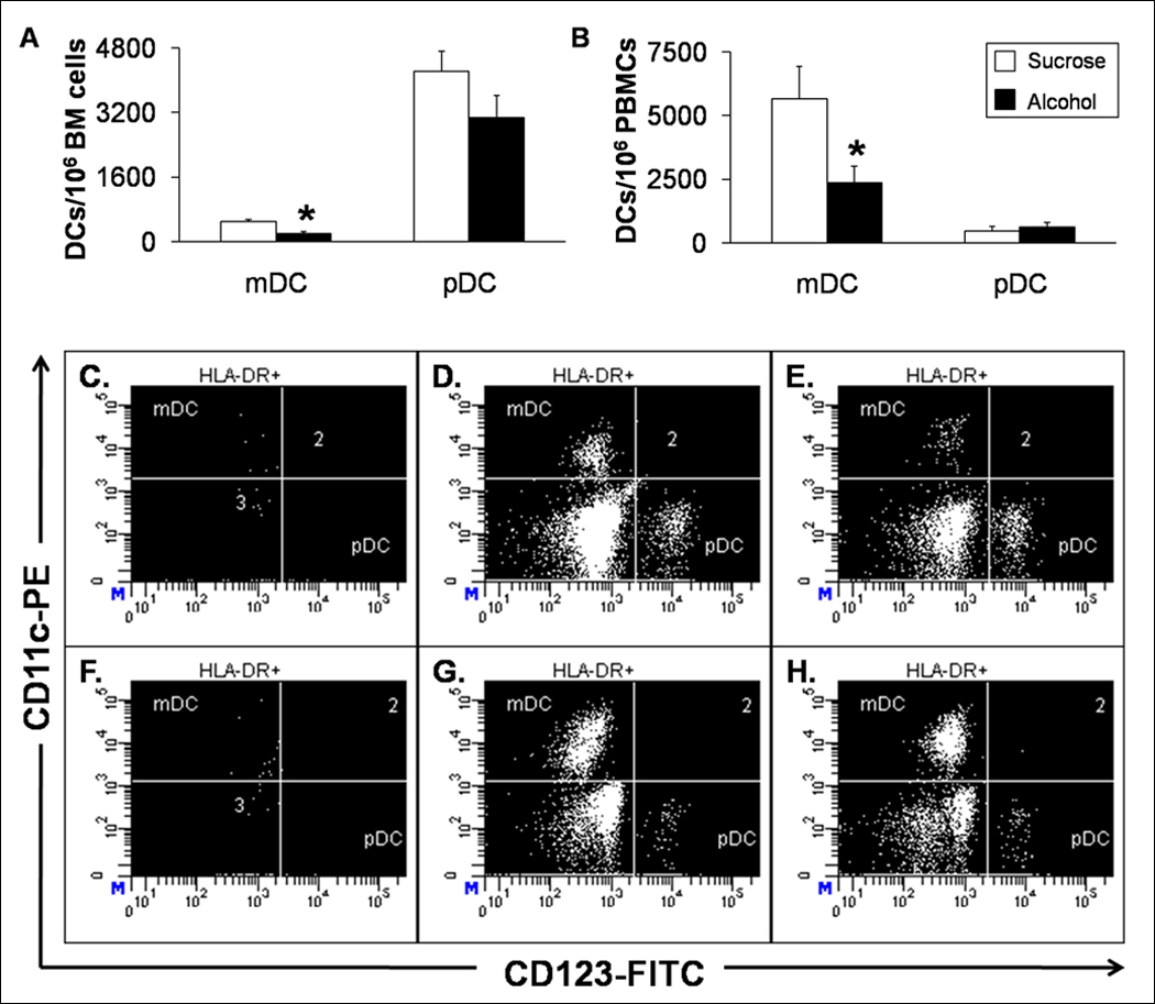
Bone Marrow and Circulating Myeloid DCs and pDCs. Alcohol decreases the absolute number of myeloid DCs in the (A) bone marrow and (B) peripheral blood in macaques. Data are mean ± SEM; N=3 for bone marrow groups; N = 8 in control and N = 7 in alcohol groups for peripheral blood; Asterisks indicate statistical difference (p < 0.05). Representative dot plots from (C–E) bone marrow and (F–H) peripheral blood are shown. The myeloid DCs are clearly diminished by alcohol (E and H) compared to sucrose-fed animals (D and G).