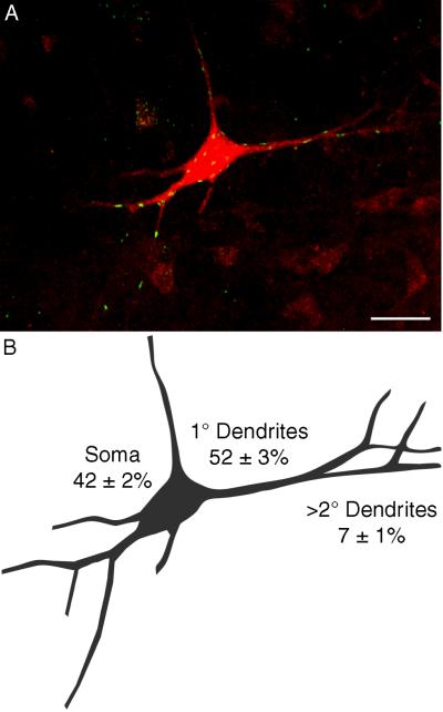
Figure 6.
Distribution of NRG1-containing synapses along motoneuron compartments.
A. Maximum intensity projection of 73 slices obtained from a 100 μm longitudinal section at C4 shows multiple NRG1-immunoreactive synapses (green) at a retrogradely labeled PhrMn (red). The background level was set to allow visualization of distal dendritic branches resulting in increased noise in the projection of this thick confocal stack. Also note that some NRG1-immunoreactive synapses may falsely appear to be within the motoneuron. The number of NRG1-expressing synaptic sites is greatest at the soma and primary dendrites. Bar: 50 μm
B. Camera lucida drawing of the motoneuron shown in A. The average (± SD) percentage of NRG1-immunoreactive synapses at the soma, primary dendrites and distal dendrites (i.e., secondary and higher order) for 26 PhrMn (n=3 animals) is indicated.

