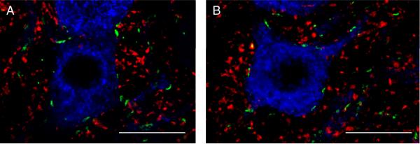
Figure 8.
Localization of NRG1 immunoreactivity at glutamatergic synapses.
A. Maximum intensity projection of two consecutive confocal slices showing a retrogradely labeled PhrMn (blue), synapses labeled by NRG1 (green), and the vesicular glutamate transporter VGLUT1 (red). There are no areas of intersection between VGLUT1- and NRG1-immunoreactivity. Bar: 20 μm.
B. Maximum intensity projection of two consecutive confocal slices showing a retrogradely labeled PhrMn (blue), synapses labeled by NRG1 (green), and VGLUT2 (red). Areas of co-localization that appear yellow designate the intersection between the VGLUT2- and NRG1-immunoreactivity. Overall, ~25% of the NRG1 immunoreactive synapses at PhrMn are associated with VGLUT2 expression. Bar: 20 μm.

