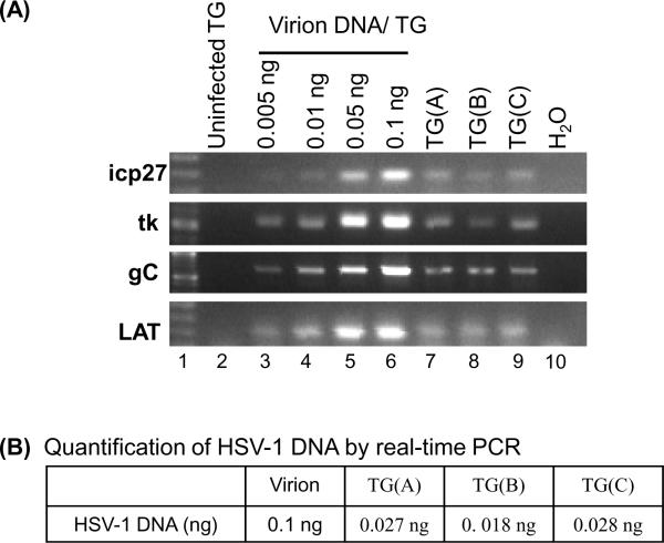
Figure 6.
HSV-1 DNA from latently infected mice is refractory to PCR. DNA was isolated from TG and the amount of HSV-1 DNA in the TG DNA sample was quantified by Southern blot hybridization as shown in Figure 3. TG(A), TG(B), and TG(C) DNA samples were isolated from TGs of mice infected with 3 × 10⁴, 3 × 10⁵, and 4 × 10⁷ PFU of HSV-1 strain F, respectively. HSV-1 DNA (1 ng) from each TG DNA sample were subjected to PCR with primers specific for HSV-1 ICP27, TK, gC, and Lat genes. PCR was also conducted using varying amounts of virion DNA reconstituted with 4.6 μg of uninfected TG DNA (lanes 3 – 6) and 4.6 μg of uninfected TG DNA (lane 2). PCR products derived from each reaction were analyzed on a 1% agarose gel, stained with ethidium bromide, and photographed. (B) Quantification of HSV-1 DNA by real-time PCR. Viral DNA (1 ng) of each sample and a range of concentrations of virion DNA standards were subjected to real-time PCR assay with primers specific for ICP0. The amount of viral DNA in each TG sample quantified by real-time PCR is shown.

