
Figure 5.
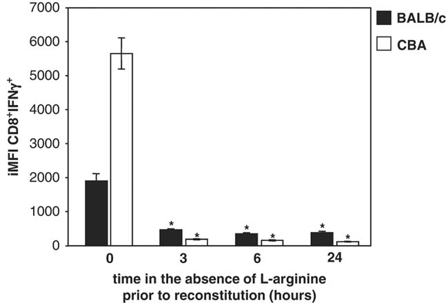
Impaired capacity of antigen-specific CD8+ T cells to express IFN-γ. Individual popliteal lymph nodes were harvested from BALB/c and CBA mice infected with L. major for 2 wk (n=4) and restimulated with L. major parasites in the absence of l-arginine. After 3, 6 or 24 h, l-arginine (400 μM) was added to the cultures. In addition, some cells were restimulated with L. major parasites in the presence (400 μM, “0 hour” group) of l-arginine. After 5 days, cells were harvested and the iMFI of cytokine-producing CD8+ T cells were determined as described in the Materials and methods. Data show mean±SD of IFN-γ+CD8+ iMFI from four individual lymph nodes/group. *p<0.05 as determined by a two-tailed Mann–Whitney test. Data are representative of two independent experiments.