Figure 5.
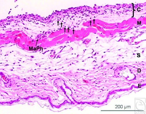
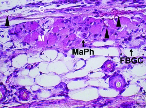
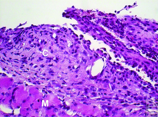
(a) The image shows the cross-section of a skin tissue sample (hematoxylin and eosin staining) that was exposed to plasma-treated collagen-coated silicone implant. (E = epidermis, D = dermis, S = subcutis, M = striated skin muscle, C = capsule). Leukocytes (arrows) penetrate the skin muscle layer (M) directed to the capsular tissue (C). Macrophages (MaPh) are evident at the capsule-muscle interface (magnification: 100-fold). (b) An extensive fibrous deposition within the implant capsule and enhanced infiltration of the capsular tissue by leukocytes were found in the untreated group (magnification: 200-fold). (c) The muscle and the fibrous capsular layer of treated implants show a high vascular infiltration (blood vessels are marked by triangles). Macrophages (MaPh) and a foreign-body giant-cell (FBGC) are detected within the muscle layer. The inflammatory cells are directed towards implant's capsule (magnification: 200-fold).


