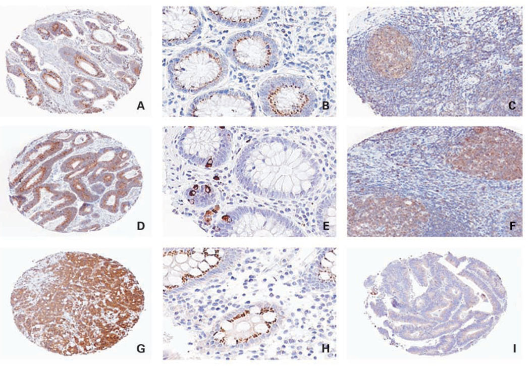
Fig. 1.
Immunohistochemical analysis of Bim, Puma, and Noxa proteins. A, colon carcinoma with elevated Bim expression in the tumor cell cytoplasm. Magnification, ×5. B, normal colonic mucosa shows Bim staining in colonic crypt epithelium. Magnification, ×20. C, tonsil shows Bim staining in lymphocytes within the germinal center (positive control). Magnification, ×10. D, colon carcinoma with elevated Puma staining in the tumor cell cytoplasm. Magnification, ×5. E, normal colonic mucosa shows cytoplasmic/perinuclear Puma staining in crypt epithelial cells. Magnification, ×20. F, tonsil serves as a positive control for Puma. Magnification, ×10. G, colon carcinoma (poorly differentiated) shows elevated Noxa staining in the tumor cell cytoplasm. Magnification, ×5. H, normal colonic mucosa with cytoplasmic Noxa staining in the crypt epithelium. Magnification, ×20.I, colon carcinoma with low-level Noxa expression. Magnification, ×5.

