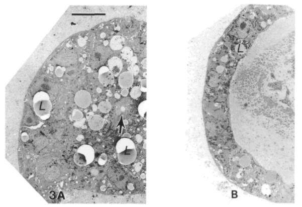
Fig. 3.
Cross-section views of secretory alveolar-like structures. After culture on EHS matrix for 8 days, cells are organized into a variety of alveolar-like structures ranging from 50-150 μm in diameter. The cells in these spheroids are polarized with their apices toward the lumen and their basal surfaces outward, contacting the basement membrane matrix. In some cases (A), the central lumen is quite small and filled with microvilli (arrow), while other (B) spheroids appear swollen with protein accumulated within their lumina. Likewise in A the cells are tall and columnar while in B the flattened cells are cuboidal. Individual cells in both of these structures show many morphological signs of secretory activity, including secretory granules, lipid droplets (L), and well-developed rough endoplasmic reticulum, although they are more prevalent in A. Bar, 10μm.

