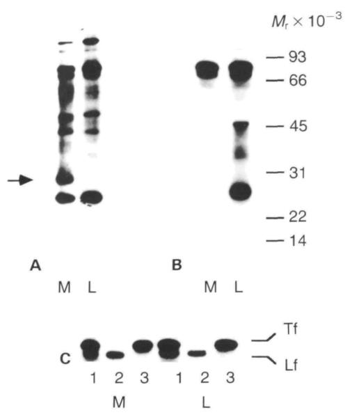
Fig. 6.
Gel electrophoretic patterns of [35S]methionine-labelled proteins secreted into the medium and the luminal compartment by primary mammary epithelial cells cultured on plastic and EHS substrata for 6 days. (A) Unprecipitated proteins. Equal volumes of medium and EGTA extract were mixed 1:1 with sample buffer and run on 12·5% PAGE. The lanes in the fluorogram show medium (M) and lumina (L) compartments and position of molecular weight standards (Mr × 10−3). Note the protein band of approximately 30×l03 Mr (arrow) that is present only in the medium of the EHS cultures. (B) Immunoprecipitated milk proteins. Equal acid-precipitable counts from medium and luminal compartments were immunoprecipitated using a broad spectrum antibody to mouse skim milk proteins. (C) Specific immunoprecipitation of transferrin (Tf) and lactoferrin (Lf) by broad spectrum milk antibody (1), lactoferrin antibody (2), and transferrin antibody (3).

