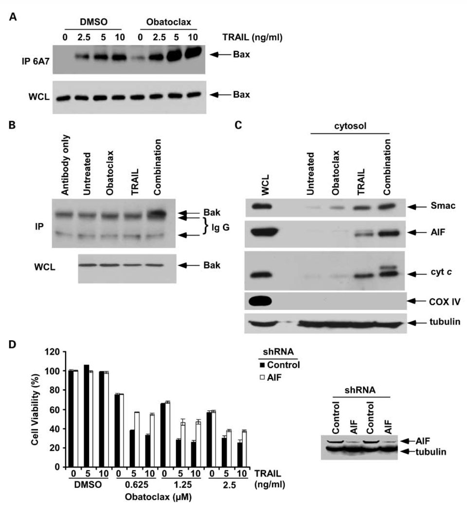
Fig. 3.
Obatoclax treatment enhances TRAIL-mediated activation of proapoptotic Bak/Bax proteins and induces cytochrome c release in PANC-1 cells. A, cells were treated with obatoclax (2.5 µmol/L),TRAIL (2.5, 5, 10 ng/mL), or their combination for 6 h. Immunoprecipitation was done using a conformation-specific antibody to Bax (mouse anti-Bax 6A7). Precipitants were then probed for Bax by immunoblotting using a rabbit polyclonal antibody. Whole-cell lysates show equal protein loading for Bax. B, cells were treated with obatoclax (2.5 µmol/L),TRAIL (10 ng/mL), or their combination for 6 h, and a Bak conformation-specific antibody was used to recognize and immunoprecipitate the activated Bak proteins. A rabbit anti-Bak polyclonal antibody and native IgG detection kit were used as primary and secondary antibodies in immunoblotting, respectively. C, immunoblot analysis of mitochondrial cytochrome c, Smac, and AIF release into the cytosol after treatment of cells with obatoclax (2.5 µmol/L),TRAIL (10 ng/mL), or their combination for 12 h. Cytochrome oxidase subunit IV was used as a mitochondrial marker. β-Tubulin was reprobed and served as a loading control. D, knockdown of AIF by lentiviral shRNA attenuates the cytotoxic effect of obatoclax combined withTRAIL, but not obatoclax alone, compared with shRNA control PANC-1 cells. Columns, mean of triplicate experiments; bars, SD.

