Figure 2.
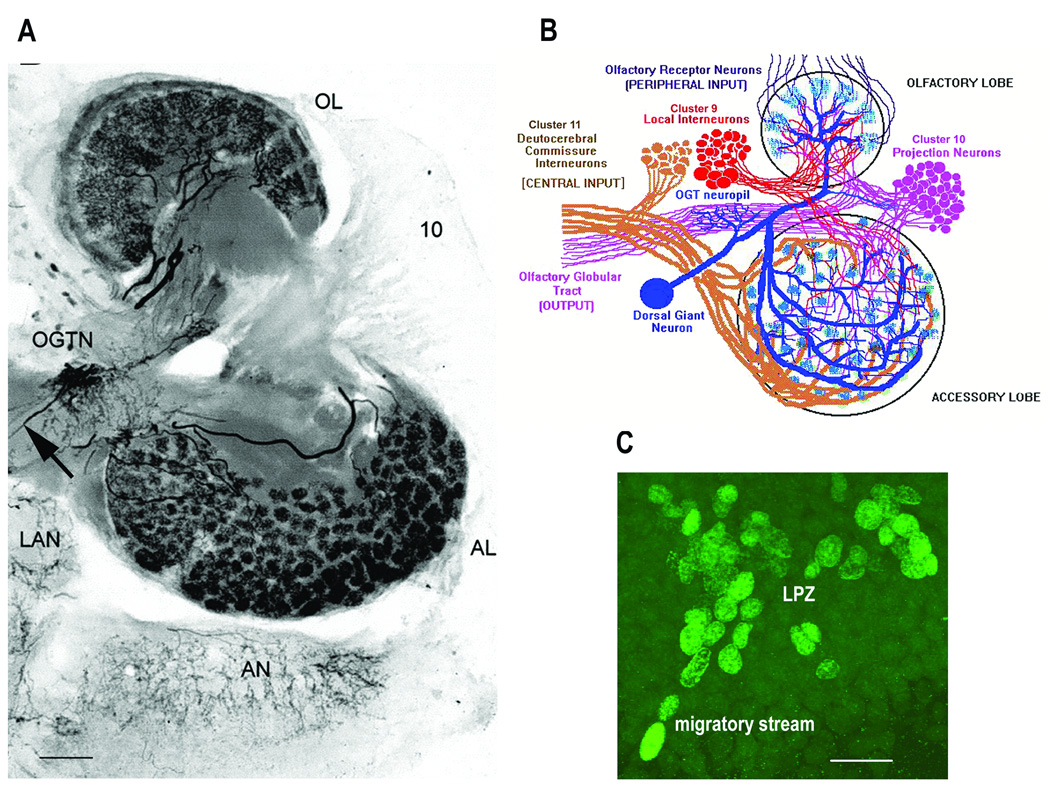
Figure 2A: Photomicrograph of a thick section (100µm) of the right side of the brain of C. destructor. Immunolabeled for serotonin the projections of the DGN into the AL and OL were revealed. The DGN is the only serotonergic neuron to project into the AL, but shares its projections into the OL with at least two other serotonergic deutocerebral neurons. The large cell body of the DGN is dorsal and out of the section plane, but the thin primary neurite that connects it with the olfactory globular tract neuropil (OGTN) before it projects into the OL and AL, can be seen (arrow). 10 – CL10 cell bodies; LAN – lateral antennular neuropil; AN – antenna two neuropil. 2B: A diagram that summarizes the circuitry of the neural elements in the OL and AL. 2C: BrdU-labeled cells at the distal end of the migratory stream and in the lateral proliferation zone (LPZ) in CL10 in a brain of C. destructor after 6 hours of perfusion with saline containing 0.5mg/ml of BrdU. Scale bars: 2A - 100µm; 2C - 20µm. (2A from Sandeman and Sandeman, 2003; 2B from Beltz and Sandeman, 2003).

