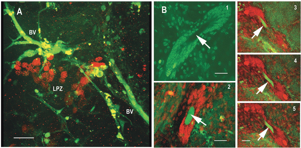
Figure 8.
Brain preparations perfused for 6 hours with saline containing BrdU and then injected with dextran dye immediately before fixation. The preparations were exposed to antibodies against BrdU, mounted and then imaged. 8A: Blood vessels (BV) filled with dextran (green) in the area around BrdU-labeled (red) cell nuclei in the proliferation zone (LPZ) of the CL10 cell bodies. The dextran was injected into the main cerebral (dorsal) artery as it entered the brain and would have passed through the capillary beds of the AL to reach these vessels in CL10. 8B: 1, the vascular cavity (arrow) in the centre of the neurogenic niche of C. destructor, surrounded by precursor cells. 2, seconds after dextran (green) is introduced into the main cerebral artery, it can be found in what we conclude is a blood vessel passing through the vascular cavity (arrow). Here the cells of the niche are red. 3,4,5, a series of optical sections taken through a niche showing a green dextran-filled blood vessel looping up through the vascular cavity (arrows). All the images in B have been brightened so that the non-specific background-labeled cells of the neurogenic niche can be seen. The niche cells in C. destructor surround the blood-filled cavity and are connected to the proliferation zones in Clusters 9 and 10 by long fibers as they are in P. clarkii and H. americanus (Sullivan et al., 2007b). Scale bars: 20µm.

