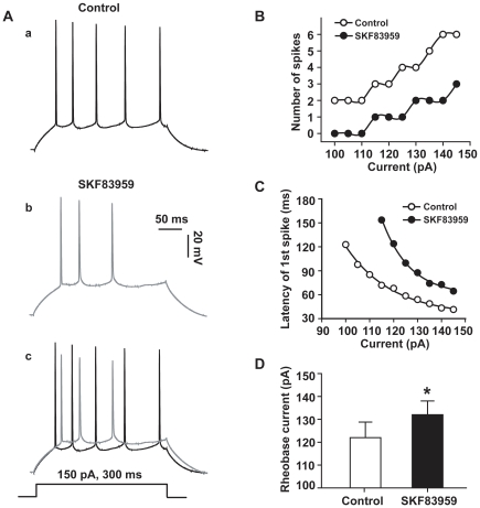
Figure 6. SKF83959 suppressed the somatic excitability of CA1 pyramidal neurons in hippocampal slices.
A. Train of action potentials of a representative neuron in response to a prolonged depolarization current pulse (150 pA, 300 ms) prior to (a) and after (b) perfusion with SKF83959 (50 µM). The resting potential was −69 mV in (a). The membrane potential was compensated by injecting steady hyperpolarizing current in (b). The two traces were superimposed at the bottom (c). B. Plot of the number of action potentials against the current intensities in another neuron. C. Plot of the latency of the first spike against the current intensities in the same neuron shown in B. The latency was defined as the time between the onset of depolarizing current pulse and the time of threshold of the first spike. D. Bar graph showing the rheobase currents measured prior to and after perfusion with SKF83959 (50 µM). *P<0.05 vs. Control.

