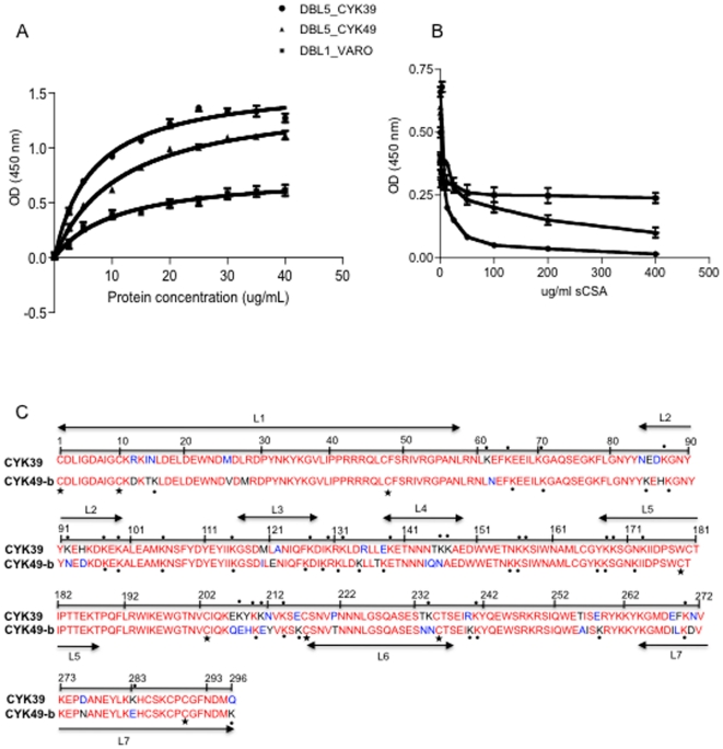
Figure 5. CSPG binding of the DBL5ε domain of the VAR2CSA from parasite isolates.
(A): Increasing concentrations of protein were added to wells coated with 5 µg/ml of CSPG. CSPG-binding of the DBL5ε_CYK39 (circle), DBL5ε_CYK49 (triangle) and the non CSA-binding VARO NTS-DBL1α domain used as control (square). Results are the means of three binding assays and the error bars indicate the standard deviation. (B) Inhibition assay. Recombinant DBL5ε variants (5 µg/ml) were pre-mixed with increasing amounts of soluble CSA 0.25–400 µg/ml, and binding to CSPG-coated plates was determined. Results are the means of three inhibition binding assays and error bars indicate the standard deviation. (C): Sequence comparison of VAR2CSA DBL5ε domains from CYK39 and CYK49. Asterisks and circles indicate respectively Cystein residues and Lysine. Conserved amino acids are shown in red and polymorphic residues in black. The 7 loops (L1–L7) identified according to Andersen P et al. [8] are underlined.

