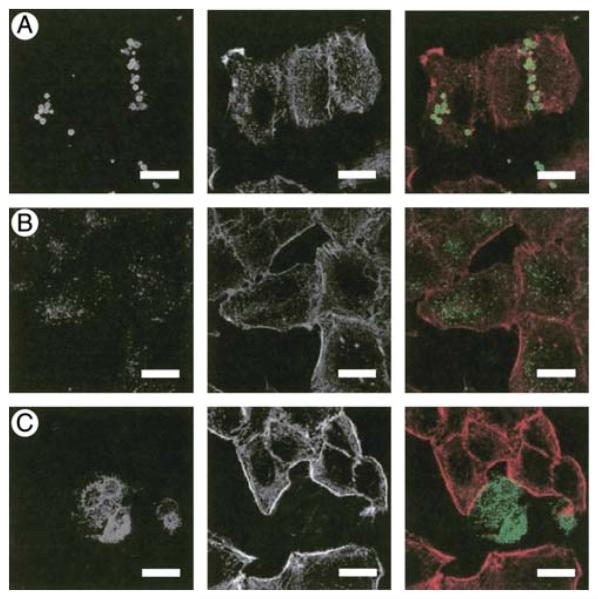
Figure 3.
Localization of LL-37B in the presence of different serum sources. A549 epithelial cells were incubated at 37°C with 10 μg/ml (2.2 μM) of LL-37B for 4 h in DMEM with (A) 10% pooled AB human serum, (B) 10% FBS, or (C) no serum. Cells were prepared for immunofluorescence assessment as described in Materials and Methods. Right panels represent a merge of LL-37B (left panels, green) and actin (middle panels, red). Bars represent 20 μm.

