
Figure 6. Transcriptional defects in IRAK-4-deficient fibroblasts.
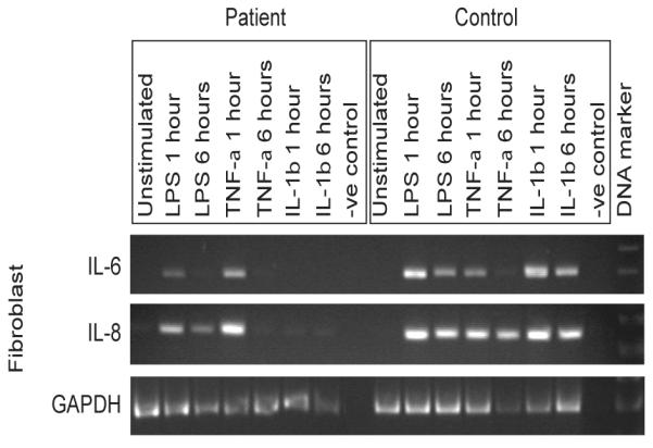
Primary dermal fibroblasts from the patient and controls were exposed to 100 ng/ml each of LPS, IL-1β, or TNF-α, or unstimulated and incubated at 37°C in 5% CO2 for 24 hr, before isolation of total RNA and semi-quantitative RT-PCR analyses, with primers designed to evaluate IL-6 and IL-8 mRNA expression, and amplification of the house-keeping gene GAPDH used as a corrective control for semi-quantitative analysis. Representative images show amplified products run on an agarose gel, with a RT-negative control, from n≥3 experimental repeats and n≥2 replicates per sample, using n=2 different normal controls.