Figs 3.
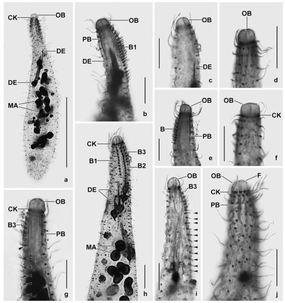
a–j. Enchelyodon alqasabi after protargol impregnation. a – body shape, infraciliature, and macronucleus pattern of a representative specimen; b–j – details of anterior body region of various specimens. Note developing extrusomes, inconspicuous pharyngeal basket, variation of oral bulge shape, and structure of dorsal brush. The dorsal brush is heterostichad, and the dikinetids of the circumoral kinety are composed of basal bodies side by side (j). Arrowheads in Fig. 3g, i mark the monokinetidal bristle tail of brush row 3. B – dorsal brush, B (1–3) – dorsal brush (rows), CK – circumoral kinety, DE – developing extrusomes, F – oral bulge fibres, MA – macronucleus nodules, OB – oral bulge, PB – pharyngeal basket. Scale bars: 50 μm (a) and 10 μm (b–j).
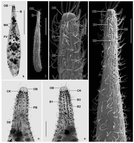
k–p. Enchelyodon alqasabi after protargol impregnation (k, n, o) and in the scanning electron microscope (l, m, p). k – dorsal view of holotype specimen showing dorsal brush and macronucleus nodules; l – general shape of a representative specimen. Arrowhead marks the end of the monokinetidal bristle tail of brush row 3; m, p – anterior body end of same specimen shown in (l). Note the structure of the dorsal brush and the cell surface covered by bacteria (arrows). Arrowheads mark the monokinetidal bristle tail of brush row 3; n, o – ventral and dorsal anterior body region of same specimen, showing the circumoral kinety with basal bodies of dikinetids side by side, the anteriorly slightly curved somatic kineties, and the short, slightly displaced anterior tail of the brush rows (arrowheads). B (1–3) – dorsal brush (rows), CK – circumoral kinety, DE – developing extrusomes, FV – food vacuole, MA – macronucleus nodules, OB – oral bulge, PB – pharyngeal basket. Scale bars: 40 μm (k, l), 10 μm (p, n, o), 5 μm (m).


