Figs 4.
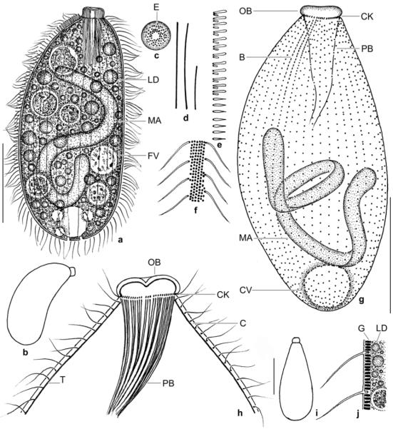
a–j. Enchelyodon nodosus from life (a–f, i, j) and after protargol impregnation (g, h). a – right side view of a representative specimen, length 150 μm; b, i – swimming specimens; c – frontal view of oral bulge. Note lack of extrusomes in bulge centre; d – two size types of extrusomes (40 μm, 20 μm); e – part of dorsal brush row 3. Note the monokinetidal tail of flame–shaped bristles; f, j – surface view and optical section showing the conspicuous cortical granulation; g – ciliary pattern of dorsal side; h – anterior body portion showing oral structures and the thick cortex separated from the cytoplasm by the darkly impregnated tela corticalis. B – dorsal brush, C – cortex, CK – circumoral kinety, CV – contractile vacuole, E – extrusomes, FV – food vacuole, G – cortical granules, LD – lipid droplets, MA – macronucleus, OB – oral bulge, PB – pharyngeal basket, T – tela corticalis. Scale bars: 50 μm (a, g) and 10 μm (h).
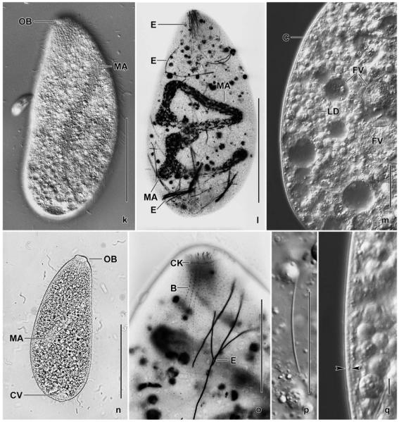
k–q. Enchelyodon nodosus from life (k, m, n, p, q) and after silver carbonate impregnation (l, o). k, n – freely motile specimens shown in interference contrast and bright field; l, o – overview and detail, showing the dorsal brush (B) and the long, deeply impregnated extrusomes; m, q – optical sections showing the thick cortex (opposed arrowheads) and the cytoplasm studded with food vacuoles, lipid droplets, and granules; p – a long extrusome. B – dorsal brush, C – cortex, CK – circumoral kinety, CV – contractile vacuole, E – extrusomes, FV – food vacuoles, LD – lipid droplets, MA – macronucleus, OB – oral bulge. Scale bars: 80 μm (k, l, n), 40 μm (m, o, p), 5 μm (q).


