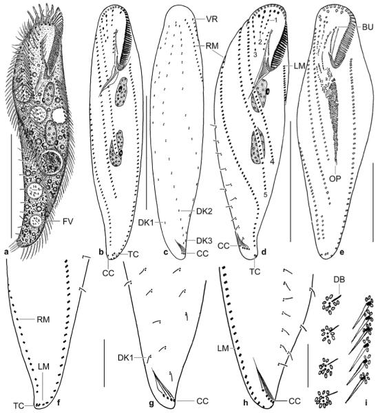
Figs 5.

a–i. Metauroleptus arabicus from life (a, i) and after protargol impregnation (b–h). a – ventral view of a representative, slightly twisted specimen, length 180 μm; b–d – ventral and dorsal view of holotype specimen (180 μm) and ventral view of a distinctly twisted paratype specimen. Both specimens have three (1–3) short and two (4, 5) long ventral cirral rows; e – ventral view of an early divider; f, g – ventral and dorsal view of posterior body region, showing the location of the transverse and caudal cirri; h – dorsal view of a distinctly twisted specimen with seven caudal cirri, of which five are recognizable; i – cortical granulation around dorsal bristles and cirri. BU – buccal cirral row, CC – caudal cirri, DB – dorsal bristles, DK 1–3 – dorsal kineties, FV – food vacuole, LM – left row of marginal cirri, OP – oral primordium, RM – right row of marginal cirri, TC – transverse cirri, VR – ventral cirral row. Scale bars: 80 μm (a–d), 50 μm (e), and 20 μm (f–h).
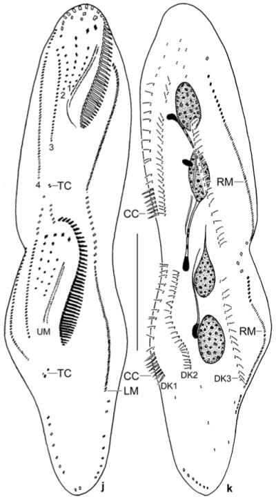
j, k. Metauroleptus arabicus, ventral and dorsal view of a late divider after protargol impregnation. Parental structures shown by contour, newly formed structures shaded black. The four (1–4) ventral cirral rows likely originated from extensions of the oral primordium, the buccal cirral row, and the two short ventral rows (cp. Fig. 5e). Note that caudal cirri are formed only in dorsal kinety 1. CC – new caudal cirri, DK 1–3 – newly formed dorsal kineties, LM, RM – newly formed left and right row of marginal cirri, TC – newly formed transverse cirri, UM – newly formed undulating membranes. Scale bar: 50 μm.

