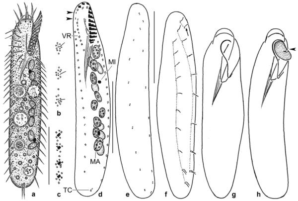
Figs 7.

a–h. Pseudohemisincirra arabica (a–f) and Keronopsis dieckmanni (g, h) from life (a, g, h) and after protargol impregnation (b–f). a – ventral view of a representative specimen, length 90 μm; b, c – cortical granulation around dorsal bristles and cirri; d, e – infraciliature of ventral and dorsal side and nuclear apparatus of holotype specimen, length 80 μm. Note lack of buccal and caudal cirri as well as the minute cirri each composed of only 2–4 cilia. Arrowheads mark frontoterminal cirri; f – dorsal view of a paratype specimen with kinetids connected by dotted lines. Arrow marks row 3 consisting of only two kinetids; g – outline of an ordinary specimen, length 250 μm; h – outline of a slender specimen just ingesting a Colpoda maupasi (arrowhead), length 220 μm. MA – macronucleus nodules in two rows one upon the other, MI – micronucleus, VR – ventral cirral row. Scale bars: 30 μm (a, d–f).
