Figs 9.

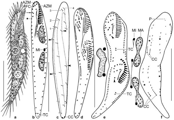
a–f. Erimophrya monostyla from life (a) and after protargol impregnation (b–f). a – ventral view of a representative specimen, length 70 μm; b, c – ventral and dorsal view of holotype specimen, length 72 μm. Arrows mark the single postoral cirrus and frontoventral cirri III/2 and IV/3, which are close together. Asterisk denotes begin of right marginal row. Arrowheads delimit gaps in dorsal bristle rows 1 and 3. Note that this specimen has only one micronucleus; d – cirral pattern and nuclear apparatus of an early divider, showing that only five (numerals) fronto-ventral- -transverse cirral anlagen streaks are produced; e, f – ventral and dorsal view of a late divider, showing that two transverse and two caudal cirri are generated. Arrowheads mark the postoral cirrus, which is migrating posteriorly. Asterisks mark gaps in dorsal kineties 1 and 3 (cp. Fig. 9c). Dorsal bristle row 4 originates close to the right row of marginal cirri. The macronucleus nodules divide a second time. Parental structures shown by contour, newly formed shaded black. AZM – adoral zone of membranelles, CC – caudal cirri, FC – frontal cirri, MA – macronucleus nodule, MI – micronucleus, P – parental dorsal bristles, TC – transverse cirri, 1–4 – dorsal bristle rows, 1–5 – cirral anlagen streaks. Scale bars: 30 μm.
