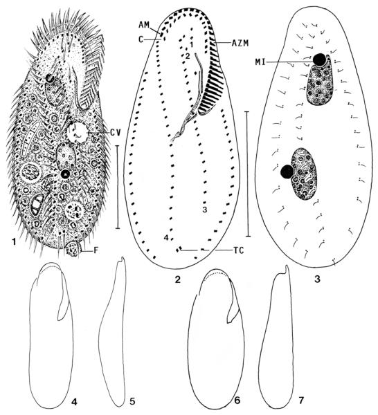
Figs 1-7.
Keronopsis schminkei, Hawaiian specimens from life (1, 4–7) and after protargol impregnation (2, 3). 1 – ventral view of a representative specimen packed with minute (3–4 μm) food vacuoles containing bacterial rods; 2, 3 – infraciliature of ventral and dorsal side and nuclear apparatus of holotype specimen. Note the transverse cirri, which are the sole difference to the genus Paraholosticha; 4, 5– ventral and lateral outline of a slender specimen; 6, 7 – ventral and lateral outline of a well-nourished specimen. AM – distalmost adoral membranelle, AZM – adoral zone of membranelles, C – coronar cirri, CV – contractile vacuole, F – faecal mass, MI – micronucleus, TC – transverse cirri, 1–4 – ventral rows. Scale bars: 40 μm.

