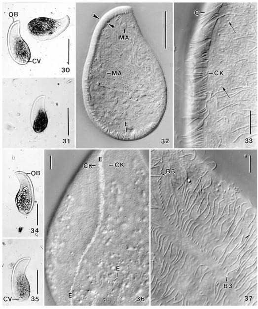
Figs 30-37.
Apobryophyllum schmidingeri, Austrian specimen from life. 30, 31, 34, 35 – overviews showing body shape and oral bulge, which is distinct due to the many extrusomes contained. The blackish areas are convex and contain food inclusions; 32 – a slightly flattened (by coverslip) specimen, showing the massive oral bulge (opposed arrowheads) and the many macronucleus nodules; 33 – mid-region of oral area, showing the densely ciliated circumoral kinety and some minute dorsal bristles (arrows); 36 – ventral view showing transverse extrusome rows in oral bulge; 37 – brush row 3 has a monokinetidal tail composed of 1.5 μm long bristles. B3 – dorsal brush row 3, C – circumoral cilia, CK – circumoral kinety, CV – contractile vacuole, E – extrusomes, MA – macronucleus nodules, OB – oral bulge. Scale bars: 100 μm (30, 31, 34, 35), 50 μm (32), 10 μm (33, 36, 37).

