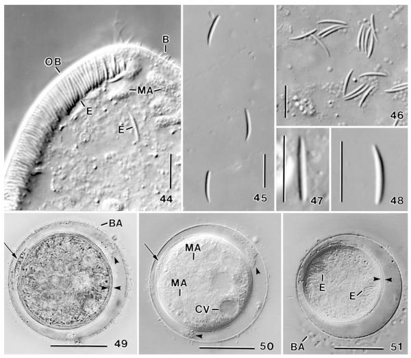
Figs 44-51.
Apobryophyllum schmidingeri, Austrian specimens from life. 44 – anterior body region showing the oral bulge studded with 8–10 μm long extrusomes (cp. Figs 45–48); 45–48 – long (type I)oral bulge extrusomes at various magnifications, showing the asymmetric shape (47, 48); 49, 50 – resting cyst in bright field and interference contrast. Arrows mark extruded material between the bipartite inner wall; opposed arrowheads denote the inner cyst wall; arrowheads mark fuzzy material between the outer and inner wall; 51 – resting cyst showing the bipartite inner wall (opposed arrowheads) and various cytoplasmic inclusions. B – dorsal brush bristles, BA – bacteria attached to the cyst wall, CV – contractile vacuole, E – extrusomes, MA – macronucleus nodules, OB – oral bulge. Scale bars: 30 μm (49–51), 10 μm (44–48).

