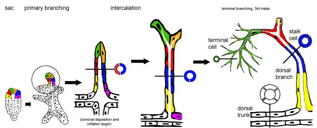
Figure 1. Three distinct tube types generated by tracheal branching morphogenesis.
From left to right: During early stages of embryogenesis, tracheal cells invaginate and form tracheal sacs composed of roughly 80 cells arranged in a polarized epithelial monolayer (“sac” schematic). Six cells are colored coded (yellow, red, green, orange, blue and magenta) to allow them to be followed over time. In response to a Branchless FGF chemoattractant cue, tip cells initiate the primary branching program, and six primary branches bud from the tracheal sac (“primary branching” schematic). Cells within the hashed circle (dorsal trunk anterior branch, to left, dorsal branch at top) are schematized at later developmental time points shown to the right. The cells of the dorsal branch are initially arranged side by side such that a cross-section view (black line) reveals the profile of two cells (red and blue) surrounding the tube lumen (“intercalation” schematic). The cells remodel their cell-cell contacts, changing neighbors (note: blue cell no longer shares a cell-cell junction with the orange or magenta cells) and intercalating to form a longer thinner tube. In a cross-sectional view, the mature dorsal branch tube is a single cell (blue) in circumference. The dorsal branch tip cells (green and orange) become specified as terminal and fusion cells, the former undergoes extensive branching during larval life, while the latter anastamoses with a fusion cell from the contra-lateral side to produce a continuous tube spanning the dorsal midline. By the end of embryogenesis, tubes of three distinct cellular architectures are present in the tracheal system. These distinct tube types are easily recognized in the third instar larvae, where terminal cells have ramified extensively, producing dozens of branched terminal tubes (“terminal branching, 3rd instar” schematic). The tubes from a single terminal cell (green) spread over areas of 100 microns or more, and are a micron or less in diameter. In cross section the tubes are revealed to be “seamless.” In contrast, the dorsal branch stalk cells (red, blue, yellow) wrap around a lumenal space and seal into a tube by forming autocellular adherins and septate junctions–represented by the single seam visible in cross section (blue). Dorsal trunk tubes are several cells (white) in circumference and the cells that compose them organize into a tube by making intercellular adherins and septate junctions –in cross section, a junctional seam is visible between all cells.

