
Figure 1. PRR in the brain of WKY and SHR.
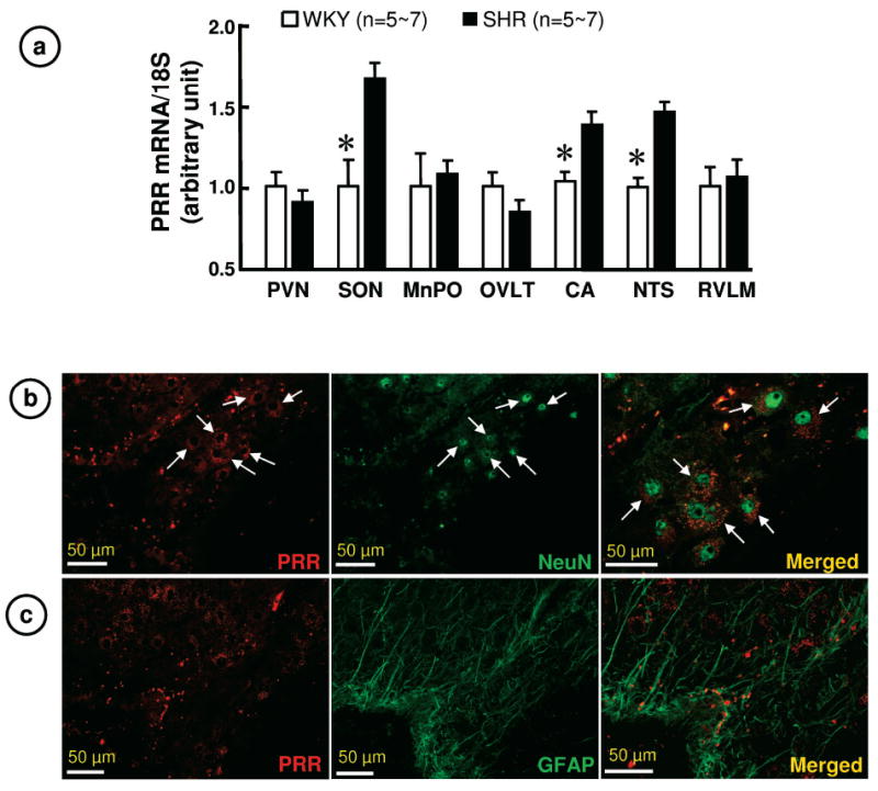
a. PRR mRNA in WKY and SHR brains. PVN: paraventricular nucleus; MnPO: median preoptic nucleus; OVLT: organum vasculosum of lamina terminals; RVLM: rostral ventrolateral medulla. *P< 0.05 vs. WKY.
b. Representative immunofluorescence micrographs using anti-PRR and anti-NeuN antibodies reveal co-localization of PRR with neurons in the SON.
c. Representative immunofluorescence micrographs using anti-PRR and anti-GFAP antibodies reveal that PRR is little present on astroglia in the SON.