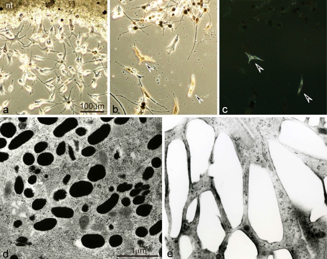
Fig. 2.
Differentiation of pigment cells from wild-type neural crest cells in culture using serum-free medium. a Differentiating melanophores and neural crest cells migrating out from the neural tube explant (nt) after 3 days in culture. b,c Differentiated melanophores and iridophores after 20 days in culture observed under transmitted light (b), or incident light (c). d,e Ultrastructure of differentiated melanophores (d) and iridophores (e) in culture. Wild-type melanophores, which differentiated first in culture, were dendritic and aggregated melanosomes (a,b). Note that melanophores also appeared on the neural tube explant (a). Wild-type iridophores, which differentiated later in culture, looked brown under transmitted light (b, arrowheads) and reflected light under incident illumination (c, arrowheads). Wild-type melanophores contained many melanosomes (d), while wild-type iridophores were filled with many rectangular reflecting platelets (e)

