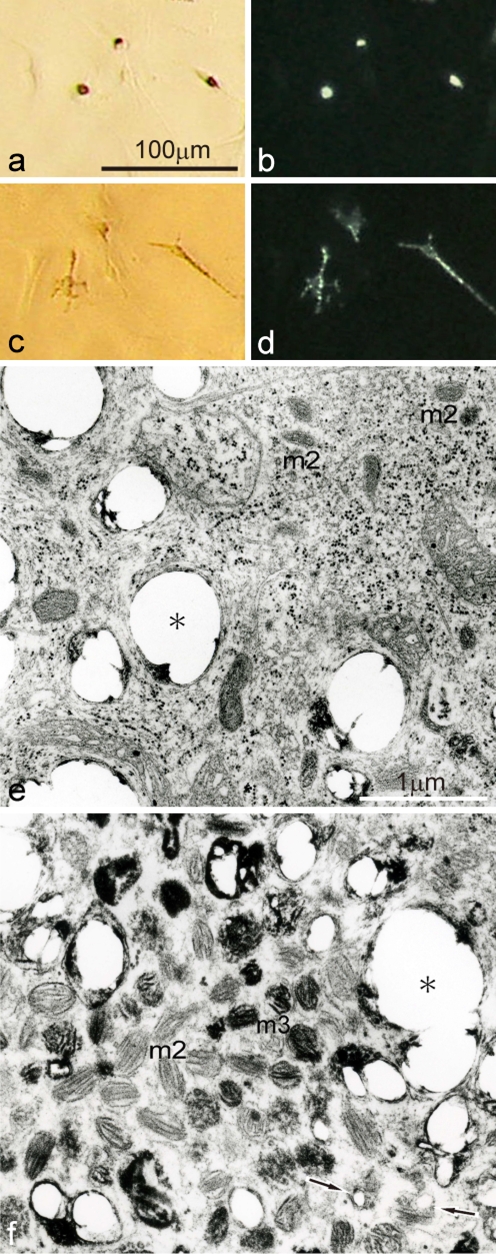
Fig. 5.
Physiological and ultrastructural characteristics of white pigment cells which were cultured from mutant tadpole tails at stage 52. a,b Cultured white pigment cells before α-MSH administration observed under transmitted light (a), or incident light (b). c,d The same fields as (a) and (b), respectively, after α-MSH administration (1 μg/ml) observed under transmitted light (c), or incident light (d). e,f Ultrastructural variation of white pigment cells in culture. White pigment cells which had a few dendrites dispersed pigment organelles in response to α-MSH (a−d). Some white pigment cells (e) contained irregular reflecting platelets (asterisk), in addition to stage II premelanosomes (m2), whose organelles were characteristic of melanophore precursors at early to middle stages of development. The other white pigment cells (f) contained irregular reflecting platelets (asterisk), in addition to stage II premelanosomes and a small number of partially melanized stage III melanosomes (m3), whose organelles were characteristic of melanophore precursors at the late stage of development. Arrows indicate premelanosomes in which reflecting platelet formation seems to be occurring

